The Complete Guide to the Muscles of the Human Neck
The head and neck muscles have many crucial tasks to perform, including the movement of the neck and head, speech, chewing and swallowing, eye movement and facial expressions. All these tasks need actions that are full of force and strength.
The face muscles are unique muscle groups in the human body, and while a majority of muscles move and connect to bones only, facial muscles mostly join your bones to the skin. The facial muscles, including the orbicularis oris and zygomaticus major, pull on your skin and allow you to make facial expressions, and move the cheeks and lips while eating and during speech.
Neck Muscle Anatomy Made Simple
Many neck or cervical muscles are superficial, which means they are very close to the skin. You can see them work beneath the skin when your body is performing actions involving the muscles. In many cases, if you put your fingers on these muscles, you can feel them work.
Below is a comprehensive list of the musculature and descriptions of their roles in supporting your head, neck and spine.
Anterior Neck Muscles :
The anterior neck muscles (anterior triangle) are located at the neck front. The front neck muscles play a significant role in swallowing and in speech, by regulating motor functions related to the larynx and the hyoid bone, a horseshoe-shaped bone found in the front of the neck below your chin. Sometimes they are called the hyoid muscles.
The anterior triangle is adjacent to the sternocleidomastoid muscle in the middle of your neck. It starts from the chin down to the manubrium, and the bottom of the mandible. The anterior of the neck has multiple muscles, which are classified in relation to the hyoid bone, which give support to muscles, which help you move the tongue or swallow.
Hyoid Muscles:
The hyoid muscles are found above and below the hyoid bone, and they move the larynx and hyoid bone
Suprahyoid Muscles:
The Suprahyoid muscles join the hyoid bone to the skull. They are found in your mouth’s floor and the larynx and help in speech. They include:
The mylohyoid starts from the mylohyoid line of your mandible and enters into the hyoid. It lifts the hyoid, the mouth floor and your tongue
- The geniohyoid starts from the inferior mental spine of your mandible and into the hyoid. It pulls the hyoid in front and above, thereby shortening the floor of your mouth.
- Stylohyoid starts from the styloid process, which is a thin and pointed piece of bone just below the ear, and into the body of the hyoid. It lifts and retracts the hyoid and elongates the mouth floor.
- The digastric has two sections, one being the anterior belly, which starts on the mandible, and the posterior belly, which starts from the temporal skull bone. The digastric enters the body and the greater hyoid horn. It works in conjunction with the infrahyoid muscles and depresses the mandible against any resistance, elevating the hyoid.

Infrahyoid Muscles:
The infrahyoid muscles are located below the hyoid bone. They provide the tongue with a base and help to keep the hyoid stable during speaking and swallowing. They include:
The omohyoid starts from the scapula’s superior border, inserting into the hyoid’s inferior border. It helps in depressing and retracting the hyoid.
The sternohyoid starts from the manubrium and the clavicle’s medial end then inserts onto the thyroid cartilage. It helps in depressing the hyoid after its elevation while swallowing.
The sternothyroid originates on the back surface of the manubrium and inserts onto the body’s inferior border and the hyoid’s greater horn. It helps in depressing the larynx and hyoid.
The thyrohyoid starts from the thyroid cartilage, inserting onto the body’s inferior border and the hyoid’s greater horn. It helps to depress the hyoid and elevating the larynx.
Lateral Neck Muscles :
The lateral muscles are situated on either side of the neck. They are majorly responsible for all side-to-side head movements and rotations.
Sternocleidomastoid Muscles (SCM)
These muscles are two of the most massive neck muscles and are located on both sides of the neck. They originate from the sternum in the chest’s center and stretch diagonally to the back of each year. The sternocleidomastoid muscles aid in the flexing of the neck and upward and sideways head rotation. Together with the scalene muscles, the sternocleidomastoid muscles help in crucial respiratory functions. They are what cause neck pain from either strains, injuries, postural problems such as tech neck, or other issues.
Sternocleidomastoid Injuries :
As mentioned above, sternocleidomastoid injuries are what cause neck pains. This is a dysfunction of the sternocleidomastoid muscle when it develops weakness and tightness with trigger points in both heads. Trigger points are tight muscle fiber bands that when compressed, are pressure sensitive. When the SCM is dysfunctional, it results in nausea, face and head pain, coryza (which exhibits cold symptoms), lacrimation or eye tearing and dizziness.
Scalene Muscles :
Previously referred to as the lateral vertebral muscles, the scalene muscles consist of six muscles on the side of the neck that come in pairs. They consist of:
- Two anterior scalene muscles
- Two middle scalene muscles
- Two posterior scalene muscles
These muscles elevate the first two rib sets and for tilting your neck sideways. Together with the sternocleidomastoids, scalene muscles help in respiration.
Scalene injuries :
Scalene myofascial pain syndrome is regional, meaning the pain starts from the neck, radiating to the arm. Tense scalene muscles trigger pain in many body parts, starting from the front and back of the arms, and the chest to the upper back. The physiotherapist must know this muscle group and the trigger points for them to effectively solve upper back, shoulder chest, hand and wrist pains.
With chest pains, these trigger point pains may lead to breathing difficulties, making one feel like they are getting a cardiac arrest. The trigger points may be caused by:
- Whiplash injuries
- Coughing excessively
- Lifting or pulling with your arms level to your waist
- Struggling to breathe, especially asthma patients
- Sleeping on the stomach with head turned sideways
- Working for an extended period with head turned sideways
- Wearing tight tie or collar
- Posterior Neck Muscles
Whiplash :
Whiplash injury is very prevalent in rear collisions. It occurs when your head is forcefully thrown forward and backward, which strains the muscles and ligaments of the neck. In most whiplash cases, the longus colli becomes weak, thus overworking the SCM to compensate for the weakness. Muscle/fascia or Myofacial pain caused by whiplash can affect the SCM for many years if not adequately addressed.
Posterior Neck Muscles
The posterior neck muscles are located at the back of your neck, and they help to stabilize the vertebral column and help with intricate head movements. The posterior triangle is a significant muscle section. It extends from behind your ear to the top of your shoulder, on either side of your neck. The muscles in this triangle are the anterior, middle and posterior scalene muscles, and work in elevating the first rib bone.
The Splenius Muscles :
These refer to the two muscle groups that originate from the back of the neck, or the splenius capitis muscles and splenius cervicis muscles. The function of these splenius muscles is to extend, flex, tilt and rotate your neck. Any injury to the splenius muscles may result in sharp neck pain, headaches, shoulder and facial pain.
Splenius Capitis and Cervicis Injury
The Splenius capitis and cervicis syndrome is a condition where the splenius capitis and cervicis muscles are injured and cause pain in the head, neck and other upper extremities. Splenius capitis syndrome may be caused by:
- Car accidents
- Poor posture
- Falls
- Sports collisions
- Direct trauma
The symptoms may include light sensitivity, nausea, rear head pain, temple headache, pain under or above the eye, and neck arm, or shoulder pain.
Suboccipital Triangle
The suboccipital triangle is a group of four small muscles that are situated at the back of the junction where the neck and head connect, under the occipital bone. The muscles in the suboccipital triangle include the rectus capitis posterior major, rectus capitis posterior major, obliquus capitis inferior and the obliquus capitis superior. These three muscles help with fine motor functions, such as small head movements.
Rectus Capitis Posterior Major :
The rectus capitis posterior major is the largest of the rectus capitis muscles and is laterally located to the rectus capitis posterior minor. It originates from the spinous process and inserts laterally onto the occipital bone’s inferior nuchal line. It helps in head rotation and extension.
Rectus Capitis Posterior Minor :
The rectus capitis posterior minor is the centermost of the suboccipital muscles and is responsible for the extension of the head. Connective tissue bridges the posterior minor and the dura mater, which is the outer meringes membrane, and may be responsible for cervicogenic headaches.
Obliquus Capitis Superior :
This muscle is the most inferior in position among the suboccipital muscles. It is the only capitis muscle with no cranium attachment. It originates from the vertebra spinal process, and it helps in head extension and rotation.
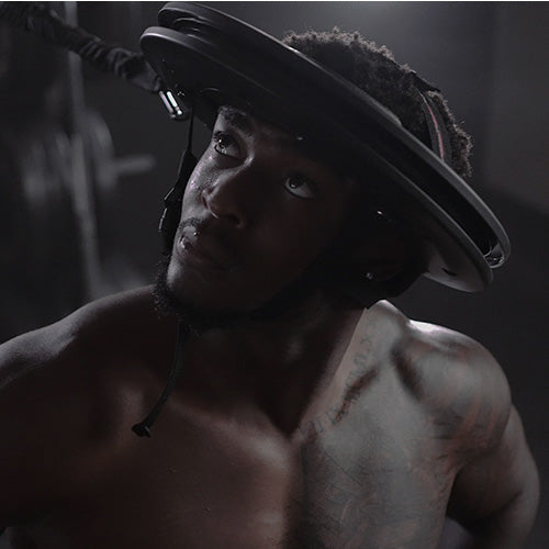
Neck Strength Training
Athletes trying to avoid neck injuries from contact sports are not the only ones who benefit from neck strength training. In our everyday lives, we encounter situations, which are far more dangerous than a punch, tackle, car accident or fall. The worst part is, if these things happen, we are not prepared for them. We may think twice about combat sports such as an NFL tackle, but do not think twice about hopping into a car and driving at 65mph.
An NFL tackle potentially produces 1,600lbs of force, while a person weighing 15—200lbs and driving at 65mph without a seatbelt can experience 160-215,000lbs of force upon impact. With a belt, the 215,000lbs of force reduces to 32,000 to 43,000 lbs. of force upon impact.
Your neck’s musculature is your head, neck and spine’s support. The Iron Neck predominantly trains your neck’s musculature isometrically. This means the musculature has no change in your overall muscle length. The neck musculature is responsible for keeping the head, neck and spine from accelerating and rotating aggressively, much as a seatbelt does.
According to Dr Uzma, a leading neurologist in concussion research, neck strengthening is believed to be the best way to reduce a concussion by preventing the rapid extension and flexion of the head, which could theoretically reduce any axonal (head, neck, spine) injuries.
Neck Strengthening Exercises
Here are some neck strengthening exercises you can do at home:
Extension and Flexion
Look upwards and extend your neck, so the tip of your nose points to the ceiling. Flex your neck so that your chin touches your collarbone’s middle. Repeat about 15 times for each side.
Lateral Flexion
Flex your neck from side to side and bring your ear as close as you can to your shoulder. Return to center and repeat on the opposite side. Do this 15 times for each side.
Wall Presses
Stand with your head and back against the wall, and pull your shoulders back. Press the back of your head against the wall, holding for one second. Relax and repeat for about 15 times.
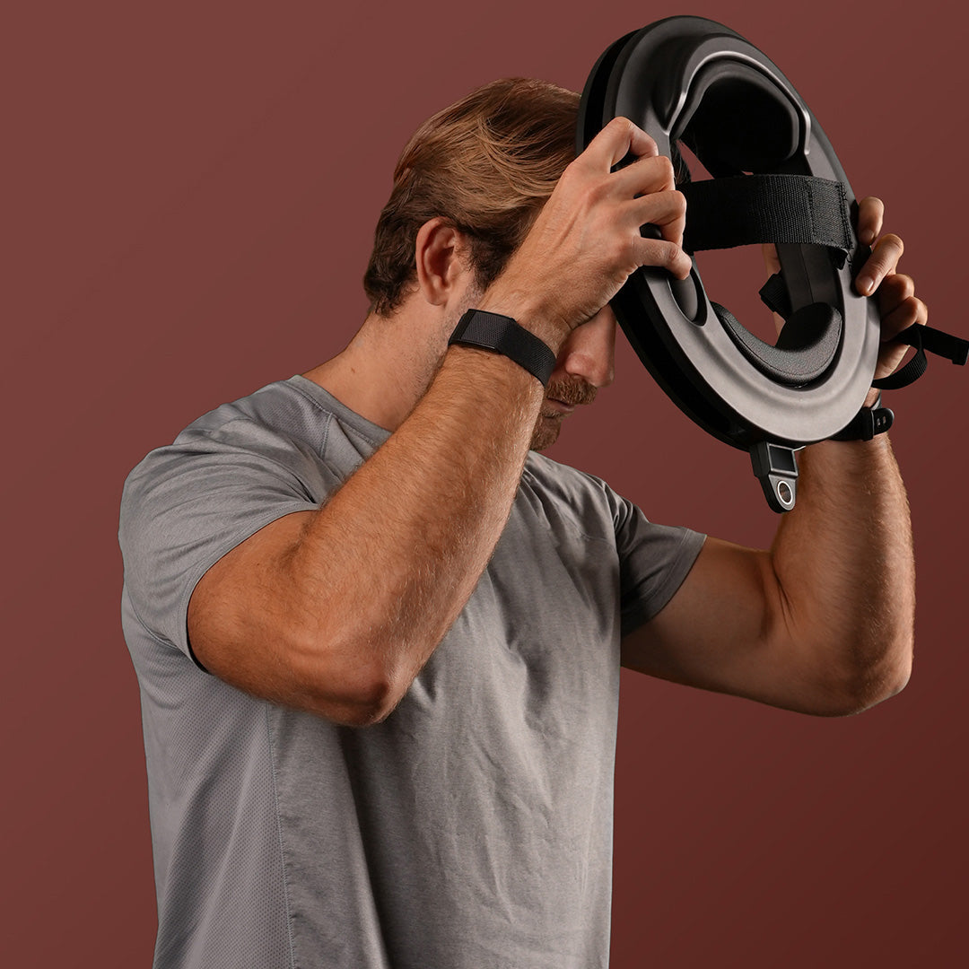
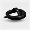
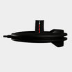
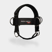
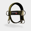
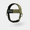
Use Iron Neck
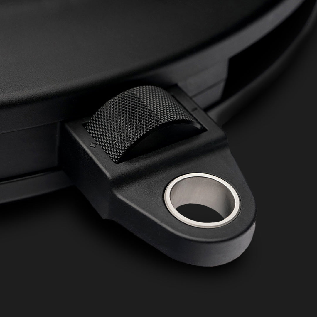
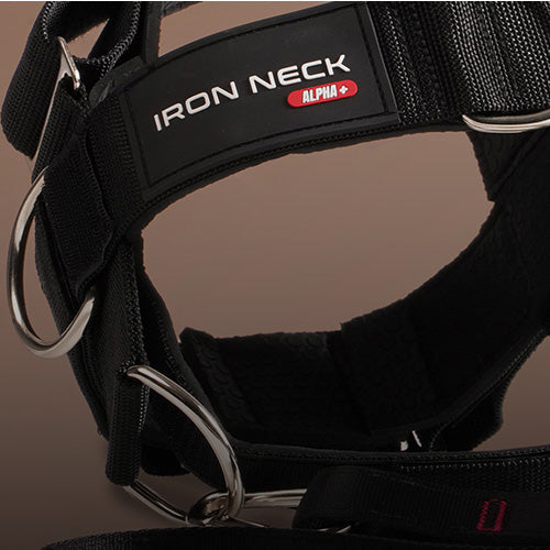
The Iron Neck is a device worn on the head and has a fitting system that includes adjustable head straps and an inflation system to hold the device in place. It works via linear and rotational resistance. Linear resistance is applied using a resistance band, which increases difficulty as the band stretches out, performance trainer, cable pulley etc. Rotational resistance is applied using a disc-brake system, which is adjustable in some iron neck models.
Conclusion
The neck is, head muscles perform myriad tasks, and injury in one muscle can affect various body parts as the muscles are intertwined. Neck injuries are prevalent, especially among car accident victims and people who engage in combat sports. To be safe, whether you play combat sports on not, it is crucial to take up neck strengthening as a precaution. This can be done via simple exercises or using an Iron Neck device.